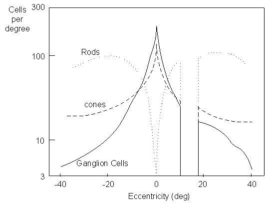
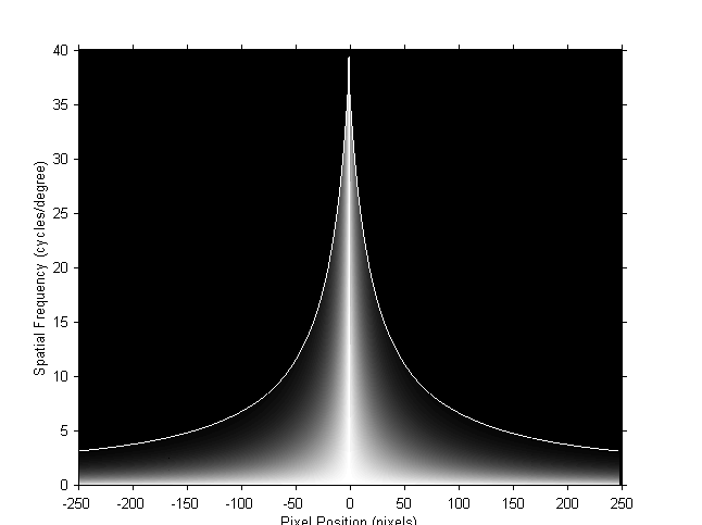
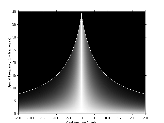
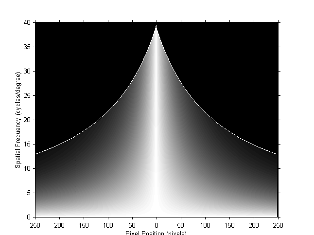
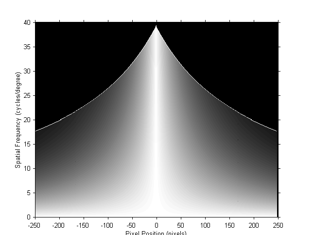
Human Visual Foveation Model The photoreceptors (cones and rods) and ganglion cells are non-uniformly distributed in the retina in the human eye as shown in the left figure below. The density of cone receptors and ganglion cells play important roles in determining the ability of our eyes to resolve what we see. Spatially, the resolution has the highest value at the point of the fovea and drops rapidly away from that point as a function of eccentricity. When a human observer gazes at a point, a variable resolution image is transmitted through the front visual channel into the high level processing units in the human brain. The region around the point of fixation (or foveation point) is projected into the fovea, sampled with the highest density, and perceived with the highest sensitivity. The right image below simulates the foveation process of the human eye.
At a certain eccentricity, the highest spatial frequency that can
be resolved by the human eye without aliasing is limited by the
corresponding sampling density in the human visual system. This
feature frequency is usually called the local cutoff frequency.
|